
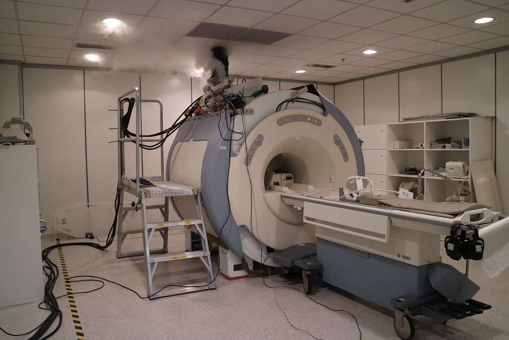
Magnetic resonance imaging (MRI) uses strong magnetic fields, radiofrequency waves, and computer processing to produce detailed images of internal body structures. Unlike X-rays and CT scans, MRI does not use ionizing radiation, and examination times vary depending on the area scanned. Radiologists examine these images to identify abnormalities affecting the brain, spine, joints, and internal organs.
Detecting a Range of Conditions
Magnetic resonance imaging provides detailed views of internal structures, helping physicians investigate symptoms and evaluate abnormalities throughout the body. MRI scans reveal changes in soft tissues, including the brain, spinal cord, muscles, ligaments, and internal organs. Because imaging needs vary, physicians select specific MRI examinations based on symptoms, medical history, and suspected conditions. Common conditions evaluated through MRI include:
- Brain and spinal abnormalities
- Joint and cartilage injuries
- Liver and abdominal disorders
- Spinal disc problems
- Blood vessel abnormalities
Preparing for a Scan Appointment
Preparing for MRI involves following safety instructions and removing objects that could interfere with the scanner. Patients typically change into a gown and remove jewelry, watches, and other metallic accessories before entering the examination room. Because MRI uses a powerful magnetic field, patients must inform staff about pacemakers, implanted devices, and other metal-containing objects.
During the scan, patients remain still while the machine produces loud tapping or knocking sounds. Many imaging facilities provide earplugs, headphones, pillows, or blankets to support patient comfort throughout the examination. Patients who experience discomfort or anxiety may communicate with the technologist through an intercom or signaling device. The technologist monitors the examination and provides instructions when adjustments or additional imaging sequences are needed.
Receiving Scan Results
MRI scan duration varies depending on the body area examined, imaging sequences, and whether contrast material is required. Some examinations take approximately twenty minutes, while more detailed scans require additional time. After the examination, a radiologist reviews the images and prepares a written report describing the findings. The report is then sent to the referring healthcare provider for review.
Referring providers use MRI findings alongside medical history, symptoms, and physical examination results to guide further care. Because reporting times vary between facilities, patients receive results according to their provider’s established process. MRI findings may support several aspects of medical evaluation, including:
- Identifying structural abnormalities in organs, joints, and soft tissues.
- Evaluating suspected injuries involving muscles, ligaments, cartilage, or spinal structures.
- Investigating unexplained symptoms that require additional diagnostic information.
- Monitoring existing conditions to assess changes over time.
- Planning further treatment based on imaging findings and clinical evaluations.
Schedule Your Magnetic Resonance Imaging Appointment
Magnetic resonance imaging uses magnetic fields and radiofrequency waves to produce detailed images of internal body structures without ionizing radiation. MRI examinations help healthcare providers evaluate injuries, identify abnormalities, and monitor certain medical conditions. Before the scan, patients receive instructions about removing metal objects, disclosing implanted devices, and remaining still during imaging. Because examination requirements vary, radiology specialists explain preparation steps, scan duration, and the process for receiving results. Schedule a consultation with a radiology specialist today to discuss your imaging needs and learn what to expect during an MRI.